Wednesday, September 9, 2015

Early data of the REVERSE-AD trial: 


N=90, 184 sites, 35 countries
Study design: prospective cohort study;
Disclosures: funded by Boehringer Ingelhelm

Inclusion Criteria: 
- People over the age of 18
- Group A: "Overt, uncontrollable or life-threatening bleeding that was judged by the treating clinician to require a reversal agent" (n=51; 18 ICH, 20 GIB, 9 trauma, 11 other)
- Group B: "Those who required surgery or other invasive procedures that could not be delayed for at least 8 hours an for which normal hemostasis was required" (n=39; Bone fractures 8 Acute cholecystitis 5 Acute renal insufficiency, catheter placement 4 Acute appendicitis 3 Joint/wound infection 3 Abscess (suprapubic, scrotal) 2 .... no neurosurgical procedures)

Primary endpoint: 
- Maximum % reversal of dabigatran as determined by dilute thrombin time or ecarin clotting time measurements

Secondary endpoints: 
- proportion of patients with complete normalization of dilute thrombin time or ecarin clotting time in first 4 hours
- Reduction in concentration of unbound dabigatran in the blood.
- Group A: Severity of bleeding (by scales - ISTH, GUSTO, modified rankin)
- Group B: h"hemostasis during the intervention was classified by the physician as normal, or as mildly, moderately, or severely abnormal"

Results: 


All of the figures basically look like this, where the lab values immediately normalize after administration of the sample 

Clinical results? 
Group A
- Of the 18 people with ICH, time to cessation of bleeding could not be ascertained in 5. Overall, the time to cessation of bleeding could not be ascertained in 13 of 51.
- Of the remaining 33, "the median investigator reported time to the cessation of bleeding was 11.4 hours"

Group B
- "normal intraoperative hemostasis was reported in 33 of 36"

Thrombotic Events:
- 5 out of 90 people
- DVT x 3, PE x2, stroke x1, NSTEMI x1

Interesting sidepoint for neurosurgeons
- "the proportion of patients with intracranial bleeding was higher among the 11 patients who had normal results on the clotting tests at baseline than among the 40 who had elevated results at baseline"
- So basically, the people who were LESS anticoagulated had more head bleeds.....

Comments: 
- Clinical endpoints are terrible - what the heck does "the median investigator reported time to the cessation of bleeding was 11.4 hours" even mean? Who is making this call? And by what metric? Is that by CT? Exam? Only 1/3 of the cases were cranial -- it was a potpourri of etiologies. Also, if the reversal happened immediately, why did it take 11 hours to stop bleeding? Is this just people getting a follow up CT scan 6 or 12 or 24 hours afterwards and seeing stability?
- No neurosurgical cases in the B group.
- Non-randomized, non-blinded, no control group


Friday, August 14, 2015

PSEUDOTUMOR 

Etiology: unknown, but there are many theories: 
- Venous sinus obstruction/stenosis, leading to back pressure in CSF -- however there's an unclear chicken-and-egg problem here, because increase in ICP will lead to compression of venous sinuses. There have been some small studies that LP's lead to decompression of venous sinuses, also some small studies demonstrating resolution of pseudotumor symptoms with sinus stents
- Microthrombi occlusion of arachnoid granulations
- Vitamin A toxicity - reducing absorption of CSF - some reports of associations between retinols and pseduotumor
- Increased intraabdominal/intrathoracic pressure leading to reduced drainage of venous blood and thus of CSF - helps to explain the obesity connection, but not necessarily the gender discrepancy, or why there isn't necessarily an association between pseduotumor and pregnancy
- Some theories about increased CSF production - however lesions that make more csf (like cpp) tend to cause hydrocephalus

Clinical presentation
- Headaches - typically severe (disproportionately so) and may present in any location with any quality- may resemble migraines or tension type HA, nor not.
- Visual loss (from papilledema) - typically long-standing, insidious onset, although a small % of people will have malignant/rapidly progressive papilledema that decompensates quickly. Typically people will lose visual fields before they lose visual acuity.
- Pulsatile tinnitus "rushing of water or wind"
- Transient visual events - vision loss, flashes, diplopia, etc.
- Typically obese women of childbearing age (20-40) - so much so that some people are reluctant to diagnose pseudotumor in people over the age of 40.
- On exam - papilledema, 6th nerve palsy, visual field cuts. Pearl -- in someone with bad enough papilledema to cause significant visual loss, you should have a 6th nerve deficit. If you don't, suspect another etiology - like optic neuritis. Papilledema can be u/l or b/l.

Workup 
- MRI to r/o secondary cause of increased intracranial pressure, like mass effect from tumor or bleed, - MRV to r/o venous sinus thrombosis
- LP to look at OP
- Measurement of systemic BP- malignant HTN can lead to optic neuropathy that resembles

Treatment
- First line - weight loss
- First line - diamox - side effects: kidney stones, metallic taste in mouth; diamox makes you lose weight, going off of it can cause it to come back. Mildly contraindicated in pregnancy.
- Second line drugs - topamax, lasix (doesn't really work)
- For severe papilledema leading to vision loss - optic sheath fenestration
- Surgical shunts - VP or LP. LP shunts obstruct like crazy, VP shunts do slightly better, but its controversial how well either works.
- Venous sinus stents - must get diagnostic angio first and have evidence of sinus stenosis; small case series seem to show good outcomes, but this is still a new and controversial procedure.

Outcomes
- It's an unfortunate reality that for most patients, the headaches do not get significantly better no matter what you do - all the medications, gastric bypass, shunts, etc. Thus don't go around shunting everyone because you probably won't help them, especially if they don't have papilledema
- People with evidence of papilledema on exam are the most likely to benefit from invasive treatment.

Sunday, August 2, 2015


Fontanelle closure: 
- occipital 1-2 mos, may even been closed at birth for some
- antero-lateral: 3 mos
- posterio-lateral: 1 year
- anterior: 9 most to 2 years.

Suture fusion 
- metopic suture typically fuses around 2 years -- all of the other sutures fuse in adulthood, starting in the 20s and continuing for the rest of life-- there is interesting data out there (like this paper) on the use of degree of suture fusion for determining age in forensics cases.

Head circumference: 
- Birth ~ 35 cm
- 3 mos ~ 40 cm
- 9 mos ~ 45 cm
- 3 year ~ 50 cm
- 9 year ~ 55 cm


Friday, July 31, 2015

Pediatric Spinal Dysraphisms 


Meningocele: 
- Protrusion of meninges filled with CSF, NO spinal cord tissue within
- Associated with hydrocephalus

Myelomeningocele: 
- Associated with hydrocephalus, chiari II (almost all), syrinx, lipomas, dermoid/epithelioid cysts, ventral and vertebral anomalies, diastematomyelia ("split cord" malformations)
- Kids who have myelo and chiari II who have hydrocephalus and fail their shunts tend to present with mild symptoms, and then clinically deteriorate very rapidly into coma and death. Perhaps the presence of the chiari II makes for less space in the posterior fossa and thus less room for progression of symptoms before acute decompensation.
- Chiari II associated with learning disability / decreased cognitive functioning, although the vast majority will be within the range of normal intelligence
- 70% will have bowel or bladder incontinence.
- Tx: prophylactic antibiotics until closure of neural tube defect to prevent infection, neurosurgical closure within 72 hours of life -- approximate edges of neural placode to create a tube.

A side note on hydrocephalus and shunt failure: its not a question of absolutes but relatives; ventriculomegaly does not have to be present, its more a function of deviation from baseline. Kids with shunts in typically have slit or very small ventricles, so a shunted kid who presents with "normal" ventricles who previously had slit ventricles may be in acute shunt failure. Likewise a kid with a relatively moderate headache who never has headaches and has had progressively worsening pain is concerning, even if the pain is not bad on an absolute scale.

Both brainstem compression or acute hydrocephalus (from shunt failure) can occur in these kids, and they can present with similar symptoms -- make sure to r/o hydrocephalus before doing a suboccipital decompression, because that in the setting of hydro can cause hernation.

Tuesday, July 28, 2015

Miscellaneous facts about peds neurology: 

First line drugs for focal seizures that generalize 
- Keppra - clean drug, no interactions, nice side effect profile
- Lamotrigine - also pretty clean, few side effects - can't be loaded - must be titrated up slowly to avoid SJS
- Oxcarbazepine - very effective, can be loaded - can cause aggression like keppra

Generalized epilepsy 
- more likely to have EEG changes at baseline between ictal events - i.e. a negative routine EEG is more likely to be expected with focal rather than generalized epilepsy

EEG
- Blinking lights tends to trigger JME
- Hyperventilation brings out absence seizures
- Indications for long term EEG - management guidance in status (spot EEG is sufficient for diagnosis - if someone is really in status, you'll see it immediately), seizures that are spaced far apart and you want to catch something.
- Key to diagnosis of seizures is rhythmicity - when your EEG looks like an EKG, you're in trouble

DDx for neonatal seizures : 
- Presenting anytime: hypoglycemia, infection, bleeding
- Presenting immediately at birth: hypoxia (~80%), withdrawal from maternal drugs
- Presenting near the first week: "Fifth day fits" - benign familial neonatal seizures, commonly inherited in families (KCNQ2 mutation), severe inherited metabolic diseases
- Presenting after first week: inherited metabolic (inborn errors of metabolism, storage diseases, etc) - tend to occur after the infant starts eating

Neonatal seizures rarely present as grand-mal/tonic clonic seizures - if these movements occur, they are more likely something else. Neonatal seizures tend to be more subtle, and may even present as isolated disturbances to vital signs (tachycardia, apena, etc)

Phenobarb and phenytoin are often used to treat neonatal epilepsy. Neither are very effective (30-40% range) and they both have terrible side effect profiles, but have been historically used and there's more information on them than other drugs -- its almost impossible to get IRB/ethics clearance to do research on neonates, so we can't do any research on newer drugs that are probably better and more effective and less toxic, so we never have any data on them, and so we have to keep using these old, crappy drugs.

More about phenytoin: 
- first order clearance until you saturate enzymes (around blood level of 20) - then becomes zero order. So the same dose that will take you from 10 to 20 might take you from 20 to 60.
- younger kids metabolize phenytoin faster, so you have to give higher doses
- phenytoin is very hard to get into solution, so you have to use a very basic solution (i.e. pH ~ 11) and that causes all sorts of problems - cardiac dysrhythmias, if it extravasates in a peripheral IV It'll cause all the skin to slough off your hand "purple stocking syndrome"
- fos-pheny is safer bc it is a prodrug that can be given in a neutral solution - its metabolized to phenytoin in your liver
- fos-pheny can be loaded faster, but its a prodrug, so in the end the onset of action is probably not that different between fospheny and pheny - and its a a lot more expensive.
- ataxia, gingival hyperplasia (bc phenytoin is excreted in saliva and evaporates, irritating gums), coarsens facial features if used for a long time, hirsutism

Primitive reflexes: 
- Moro - you drop their head down quickly, and they bring their arms together in a C, should be symmetric
- Forced head turning - they posture into a fencer stance with the ipsilateral arm extended
- Grasp - hand and foot
- Upgoing babinski
- Snout - tap closed lips, will purse
- Root - touch cheek, infant will turn towards that side and suck
- Dazzle - bright light over closed eyes, will close eyes tighter and will extinguish with repeated light

Infantile Spasms
- Treated with prednisone/ACTH - if they receive early treatment, can prevent long-term neurological damage
- Associated with tuberous sclerosis
- Very consistent timing-- you could time a watch by the regularity of the spastic events

AE Drugs that come in an IV loadable formation
- Pheny/fospheny
- Keppra
- Oxcar
- Phenobarb
- Most benzos

Monday, July 27, 2015

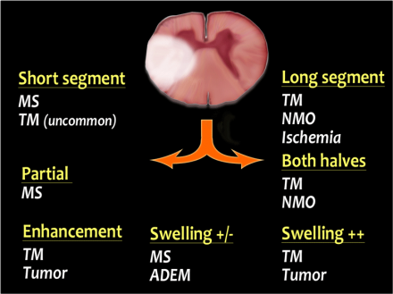
Myelopathy + a diffuse T2-hyperintense region of cord

Playing the odds 
- most likely is compression - from disc, tumor (met), trauma
- in the absence of that, most likely is demyelinating, esp MS - esp if its short segments, and there are multiple disparate lesions in random-seeming locations.
- long-segment disease - think NMO (esp if there are optic sx) or transverse myelitis (not a dz, a description of a constellation of symptoms that are caused by other things)

Transverse Myelitis etiology:
- Viral - HSV, VZV, CMV, EBV, enteroviruses (echo, coxsackie, polio), HIV, influenza, rabies, HTLV (human T cell lymphoma virus),
- Bacterial - TB, Lyme
- Autoimmune - Sarcoid, Sjogrens, Lupus
- Post vaccine - rabies, cowpox
- MS
- Paraneoplastic
- Vascular - DAF, AVM, clot of spinal aa, heroin vasculitis

Intramedullary Spine tumors
- In adults, ependymoma > astrocytoma
- In kids, astrocytoma > ependymoma
- Astrocytomas tend to look more like diffuse, +/- enhancing regions of cord with a good bit of swelling, and they tend to be associated with slowly progressive symptoms - in the context of an enhancing lesion with rapidly progressive sx and optic symptoms - think NMO, rather than astrocytoma.
- Ependymomas and Hemangioblastomas tend to look more like actual tumors
- Ependymomas often have a cap of hemosiderin at the top and bottom - from prior bleeds - and may have a cystic component

Super helpful diagrams from radiologyassistant.nl:

MS 
- Active lesions in the spine, tend to NOT enhance - in the brain, can enhance 
- If you suspect spine MS (i.e. multiple non-enhancing lesions that are small, and separate in time and space) - then get a brain MRI - if you see characteristic lesions in the corpus callosum/periventricular/pons/subcortical area, increases likelihood of MS
- 1/3 will have spinal symptoms, 95% will have spinal lesions (path dx) - irrespective of symptoms 
- 1/3 of people will have spinal MS ONLY and no brain MS 

Thursday, July 16, 2015

Decompressive Hemicraniectomy in Ischemic Stroke 

RCTs:


DESTINY
DECIMAL
HAMLET
DESTINY II
N (final) 
32
38
64
112
Year
2004-2005
2001-2005
2002-2007
2009-2013


Age range
18-60
18-55
18-60
>60
Median age
45
43 (mean)
47-50
70
NIHSS (R/L)
>18/20
>16
>16/21
>14/19
Infarct size (CT)
> 2/3 MCA territory
>1/2 MCA territory + DWI vol > 145 cm2
> 2/3 MCA territory
> 2/3 MCA territory
Median Time to surgery
24 hours
20 hours
>41 hours
28 hours
Exclusion Criteria
mRS >1, bilateral blown pupils, GCS <6, IPH, other serious illness. 
mRS>1, big contralateral infarct, big IPH 
thrombolysis w/in 12 hrs, mRS > 1, bilateral blown pupils, entire hemisphere stroke
mRS >1, bilateral blown pupils, GCS <6, IPH, other serious illness. 

Pooled results from DESTINY, DECIMAL, AND HAMLET: 


- half of the HAMLET trial pts excluded for being randomized too late (> 45 hours after symptom onset -- assuming >48 hours to treatment) 

Thoughts: 
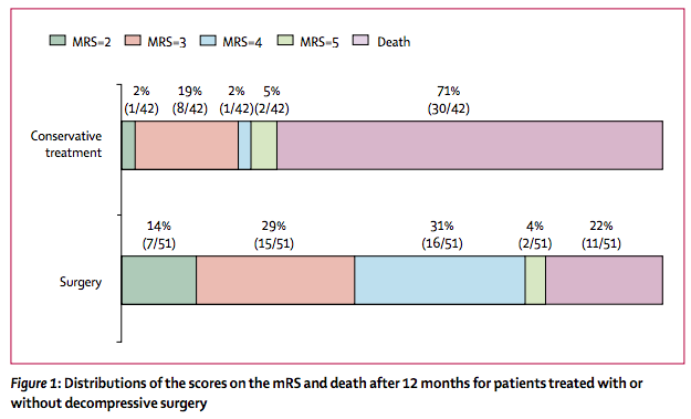
- Decompressive hemicraniectomy (DH) is associated with a significantly reduced proportion of death, but a significantly increased proportion of severe disability (mRS 4-5). 
- Incidence of very good outcome (mRS) is much higher in the surgical intervention group - 14% than the nonoperative group - 2% 


DESTINY II: