All images from this paper: http://www.ncbi.nlm.nih.gov/pubmed/17620469
Embryology: anterior pit from rathke's pouch (from mouth), posterior + infundibulum from neuroectoderm
Lesions:
Craniopharyngioma:
Germinoma
 young kids, can have pituitary/pineal synchronous lesions, homogenous, enhancing, non-cystic/heterogenous
Hamartoma
 older kids, tuber cinereum, can be pedunculated or sessile (more likely to be assoc with gelastic seizures & precocious puberty if latter). These do NOT enhance.
Dermoid Cyst
 well-circumscribed, ectodermal cysts of fat/sebaceous tissue/hair/etc. Typically T1 bright and suppress on STIR, but not as bright and not as suppressy as lipomas. Typically occur in the midline although not commonly at suprasellar. rarely enhance or have calcifications
Vs epidermoid cysts, which have the MRI appearance of dense CSF, more commonly appear off-center - esp parasellar
Vs arachnoid cysts which look just like CSF because they are
Rathke's Cleft Cyst
Hypothalamic/Chiasmatic glioma:
Ganglioglioma
Encephalitis
LCH
 LCH - has unexplained predilection for pituitary stalk and infundibulum. hypothalamic lesions enhance.
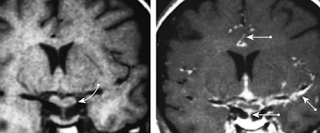
Sarcoid
nodular thickening of chiasm, stalk, infundiblum from granulomatous involvement of dura. predilection for skull base -- neurosarcoid often manifests around hypothal/pit. meningeal enhancement. can invade virchow-robin spaces.


No comments:
Post a Comment
Note: Only a member of this blog may post a comment.