Sunday, December 20, 2015


UCAS (prospective) 
- 2001-2004 in Japan 
- N=5720 patients, 6697 aneurysms (3050 : treatment before rupture @ median of 48 days, 3647: not treated before rupture) 
- Rupture rate of 0.95% per aneurysm-year ; rupture rates associated with 35% mortality, 29% mRS 3-5 

Rupture rates according to location and size:  
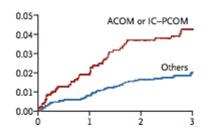
Whereas ISUIA found increased rates in all posterior circulation aneurysms, UCAS found higher rupture rates with Acomm and Pcomm (but not more with say, BTAs..... however the total number of VA/BTA aneurysms was small (see below chart)

Graphical representations of rupture rates by location and size: 


Multivariate analyses of predictors of rupture: 
*smoking status (former or current) not associated with rupture rate! 
*previous SAH not predictive of rupture - however only 3% of the cohort had ever had a SAH 

Criticisms: 
- Japanese population: which has the same incidence of aneurysm, but a higher risk of SAH compared to the rest of the world. Unclear if this data can be applied to US or european populations.
- Same selection bias as ISUIA - non-randomized data; the aneurysms believed to be high risk were all treated.


ISUIA (prospective) 
- 1991-1998
- N=4060 patients (1692 no surgery, 1917 surgery, 451 endovascular)
- Overall rupture rate -- 3% over 4.1 years of follow up; 65% mortality with ruptures

Rupture rates by location, size and presence of previous SAH*


 *All aneurysms examined in this study were unruptured, however some people in the study had a history of SAH from another source/another aneurysm. Those were designated as "group 2" --- vs "group 1", which denotes those who had no history of SAH.

Graphical representation of rupture rates by size and history of SAH: 

Multivariate model of predictors of aneurysmal rupture: 
Size: 
<7 mm (ref) 
7–12 mm, [RR] 3·3 [95% CI 1·3–8·2], p=0·01  
> 12 mm, [RR]17·0 [8·0–36·1], p<0·0001

Location:**
ICA (ref)
Basilar tip - [RR] 2·3 [1·1–4·8], p=0·025
Cavernous - [RR]  0·15 [0·04–0·64], p=0·01
Pcomm - [RR] 2·1 [1·1–4·2], p=0·02

Age: [RR] 1·007, [0·98–1·03], p=0·56 

- Morphological characteristics such as multiple lobes, presence of a daughter sac, and a family history of subarachnoid haemorrhage were not predictive of SAH 

**other locations not statistically significantly associated with rupture


Table comparing the observation vs intervention cohorts. 

Criticisms of the study: 
- Data from 1991-1998 -- before the era of widespread use of aspirin and statins, which decrease the rate of aneurysmal rupture. Also really before the era of endovascular techniques, which have significantly altered practice patterns such that the data from the people who were observed during this study is unlikely to apply to people we would observe today.
- Not randomized -- (most) everyone who was believed to be a high rupture risk underwent surgical or endovascular interventions; thus there's a selection bias for the low-risk aneurysms, thus possibly leading to an artifically lower rupture rate in the observation group.
- Selection bias of older, sicker patients --  The 5 year KM mortality in this group was 12.7% -- you would expect <1% in an age-matched control cohort. The observation group was likely enriched for poor surgical (and poor mid-90s era endovascular) candidates -- many died from cancer, heart disease, etc; About half of the patients died from intracranial hemorrhage events, many of which were NOT counted as rupture events but as censored -- patients who died from intracranial hemorrhage that could not be definitively attributed to the aneurysm were counted as censored. Another group (I can't remember which, but I think it was the TEAM group) re-analyzed the data counting all these censorship events as rupture events and found the overall annual rupture rate to be 1.2% vs 0.8% - a 50% difference although a small absolute difference.
- 32% censorship/crossover for treatment  -- enough said.
- <22% with >4 years of fu -- enough said.
- Included cavernous carotid aneurysms - which are known to have a very, very low rupture rate. 


Monday, November 30, 2015

Pretty Anatomy

Posterior choroidal vessels - lateral and medial. Lateral makes a more sharp C shape, while medial makes a curvy, swoopy "3" shape 

Lateral choroidal - makes clear C shape in lateral and AP shots. 

Really nice anatomy, ICA injection with reflux into basilar and PCAs via a large pcomm. Demonstrates a lovely anterior choroidal (orange), MHT (pink), ILT (cyan) and a mystery vessel in green that might be an enlarged vidian. 
Case study: vertebral dissection 


Young person, trauma, some C2 fractures. Concern for R vertebral artery dissection. 
Thoughts: 
- Clots respond to flow. In a case of complete occlusion leading, the clot will propagate to the nearest point of flow-- in this case, the original occlusion/dissection was at C2-3, and the occlusion is complete. We would expect it to go all the way down to the origin. It hasn't -- there are 2 reasons. One is that there might not have been enough time. Two is that there are vessels causing flow somewhere along V1 or V2 keeping the vert patent. Big muscular branches or there's sometimes a big radiculomedullary branch (analogous to artery of adamkiewicz) that comes off around C5-6 -- this is very important; occlusion of this vessel may compromise high cervical spine vascular supply. One might think, in a case where the vert is still partially filling, if we fully occlude the vert there will be a lower stroke risk -- that might be the case, but if the vert is patent because there is flow to a large radiculomedullary branch, occluding it might lead to a devastating outcome.
- Complete occlusions typically do not have to be treated with anticoagulation as they are low embolic stroke risks. Partial or recannalized verts typically do have to be anticoagulated. 

Tuesday, November 24, 2015

Weird case: 50-something with longstanding history of diplopia 


Lytic skull base lesion seems centered around petrous bone/foramen lacerum. Smooth, rounded, sclerotic (not obvious in this but clearer in other cuts) suggestive of benign, slow growing process like a vascular malformation rather than an invasive, destructive cancer like osteosarcoma (where you'd see spiculated bony edges)


Mottled appearance on thin-cut T1 GAD

Very mottled appearance on thin-cut T1 GAD 


The danger of volume averaging -- on the thicker-cut T1 GAD slices, mass appears much more homogenously enhancing.


One tiny dot of flow void/susceptibility on GRE, but otherwise nothing. 

Differential Diagnosis: 
- Trigeminal Schwannoma - although one would expect the tumor to follow the course of the trigeminal nerve, and it doesn't
- Meningioma - doesn't display bony hyperostosis, however in the literature only 25-50% of skull base meningiomas demonstrate hyperostosis, This paper (http://www.ncbi.nlm.nih.gov/pubmed/10201298) found a 51% rate and histologically showed that the vast, vast majority of cases of hyperostosis was secondary to tumor invasion into the bone. Although there were patients with tumor invasion into bone that did not display hyperostosis. 
- Hemangiopericytoma: possible given clivus bone bmarrow signal heterogeneity (which may be reactive). Per this paper (http://www.ncbi.nlm.nih.gov/pubmed/21529166), hemangiopericytoma can look very similar to meningiomas on imaging. Ways to differentiate: hemangiopericytomas frequently have serpentine flow voids, lobulated edges, mottled enhancement, erode into neighboring bone, are not surrounded by a ton of edema, and almost never have calcifications. Meningiomas typically have smooth edges, homogenous enhancements, ~20-25% are calcified. Hemangiopericytomas are also much more rare. On angio, hemangiopericytomas are usually supplied by ICA (vs ECA for meningiomas) have corkscrew vessels arising from main feeder artery, "dense, fluffy, long-lasting" tumor stain rather than sunburst blush of meningoma. 
- Mets, although no permeative bone destruction, and no known primary
- Paraganglioma: no susceptibility/flow voids
- Venous malformation -- typically these have flow voids/susceptibility, and they look different-- on T2 or T1 GAD, they look almost like halfway between an AVM and a tumor -- nests of these dilated, wormy appearing things; they are congenital and have no respect for traditional anatomic boundaries or tissue planes - can be in bone/soft tissue/etc. This tumor is more smoothly enhancing and homogenous 
- Vascular tumor - not the same thing as a vascular malformation. 

Friday, November 20, 2015

PRESSORS

Back to basics: 
- Most tissues need MAP > 60 to perfuse, brain needs CPP > 60.
- V1 is expressed in blood vessels and leads to vasoconstriction, V2 is expressed in renal ducts and leads to increased absorption of water and Na.
- Remember that the catecholamine system is mediated by cAMP
- Synthetic pressors: dobutamine, isoproterenol, phenylephrine

Epinephrine 
- at 0.01-0.1 mcg/kg/min - B1 effects predominate at these lower doses, and you get mainly inotropy/chronotropy
- 0.1-0.2 mcg/kg/min - a1 effects predominate at higher doses, and you get lots of boost to SVR
- Onset is fast, need be titrated every 2-5 mins
- uses: ACLS, inotropic support post cardiac surgery, second line for sepsis after norepi and vaso.
- problems: tachyarrhythmias, vasoconstriction of splanchnic and renal vessels, lactic acidosis, hyperglycemia to the point where people need to be on an insulin gtt.
- for anaphylaxis, you give 0.3mg IM, can repeat every 15-20 mins. If severe, can give IV epi 0.1mg slow push. (in people on b-blockers, can have paradoxical response to epi - in which case, glucagon and ipratropium - good paper in circulation on anaphylaxis)

Norepinephrine (Levophed) 
- dose ranges 0.01 to 0.2 mcg/kg/min. There is technically no upper limit but people rarely go above 0.2.
- almost all a1 activity - has very minor B1/2 activity that counteracts the reflex bradycardia so that you get net no effect on CO/HR.
- First line for sepsis and for cardiogenic shock with low SVR. Also tends to be our go-to for hypotension and vasospasm in the neuro ICU although there is approx zero data on this.
- problems: tachyarrhythmia - less than epi/dopamine at equipotent doses. Digital ischemia at high doses.
- onset in 1 min, t-1/2 5 mins, q2-5 min titration

Vasopressin 
- 0.03-0.05 units/min (0.002-0.005 units/kg/min, 1-10 units/hr)
- Binds to V1 and V2, augments SVR and MAP, has no effect on the heart.
- Uses: first line for distributed shock along with NE, variceal bleeding (in europe they use turlipressin)
- One of the advantages is that it works well at low pH - the catecholamine based pressors start to stop being effective below pH of 7.2 to 7.3
- Onset 5 min, t-1/2 10 mins.
- Problems: cardiac ischemia, can cause severe peripheral ischemia, splanchnic vasoconstriction. At very high doses, around 10x the dose we normally use, can reduce CO.
- Thought process is that in sepsis, there is a relative deficiency of vasopressin/ADH - and so 0.03 units/min replaces the endogenous supply

Phenylephrine (Neo) 
- 0.01 to 0.3 mcg/kg/min.
- Pure a1 effects. Increases SVR and MAP, reflex decrease in HR and CO (from dec HR and from afterload)
- uses: neorogenic shock, sepsis, preferred in patients who are tachycardic.
- "push dose" for procedural hypotension - 50-100 mcg every 1-2 mins prn. Anaesthesia loves this drug for intraop hypotension.
- You have to be really careful with dosing though -- a standard premixed syringe in the OR is 100mcg/mL. A bag for drip is 200mcg/mL. And if you order it from pharmacy, they will send you a vial which is 10 MG per mL, which is 100x more potent than the premixed syringes. 100x!!!

Dopamine 
- 1-3 mcg/kg/min: very selective for dopa receptors: causes renal/mesenteric vasodilation, leads to increased UOP. People used to think this dosing was "renal protective" but the trials show no change in the onset of of renal failure, no change in rate of progression to HD. Sometimes liver surgeons will use it to decrease CVP while augmenting BP.
- 3-10 mcg/kg/min: selective for B1, semi selective for D, inotrope and chronotrope
- 10-20 mcg/kg/min: selective for a1, augments SVR
uses: sepsis (rarely used except in those people who are both septic and brady or people who are really crashing and you're reaching for a 4th/5th/6th gtt), cardiogenic shock with low MAPs.
- This is one of the few pressors that people are typically comfortable giving in a peripheral line, up to doses of 10 -- so at one of the hospitals where I trained, the medicine/cards teams loved to use DA gtt on CHF-admitted-to-the-wards-for-diuresis patients when lasix alone wasn't working bc the EF was too low.
- Also easily available at most places premixed so if you're at some random hospital, you are likely to have this most easily and quickly avail.
- Problems: more tachyarrhythmias than levo at equipotent doses
- Onset in 5 minutes, half life of 2 mins.

Isoproterenol 
- Pure B1 effects. Augments CO and HR.
- Rarely used except in some heart transplant patients. As a result has become very expensive. Which means we use it even less.

Dobutamine 
- 1-20 mcg/kg/min
- Selective for B1
- Causes reflex vasodilation and decrease in MAP.
- Uses: acute decomp HF, cardiogenic shock with OK MAPs. In people who take it home in a backpack for HF, more dangerous bc the half life is 2 mins (onset is 2 mins) - so if your pump fails you're in trouble fast.

Milrinone 
- 0.1-1.0 mcg/kg/min -
- PDE3 inhibitor
- Causes vasodilation esp in pulmonary vasculature, augments inotropy
- Uses: acute decomp HF, in combo with epi after cardiac surgery. Cardiac surgeons love this drug.
- Problems: hypotension, reversible thrombocytopenia (cAMP is important in megakaryocyte differentiation - as soon as you stop the drug, the megakaryocytes can go back to producing platelets)
- Has very long half life- onset 15 mins, t-1/2 1-2 hours. Titrate slow.


SOAP II trial 


- N= 1679, multi center, randomized, double blinded trial of NE vs Dopamine
- Inclusion: <18, shock from any cause (defined as MAP<70/SBP<100 despite fluids + evidence of end-organ damage or lactic acidosis)


Bottom line: essentially no difference in survival (NE trended towards better) - way higher incidence of arrhythmia with DA


CATS Trial: 


- N=330, multi center, randomized double blind NE + dobutamine vs epi
- Inclusion: septic shock (infection + SIRS + organ hypoperfusion) with SBP/MAP requiring pressors.


Bottom line: no difference


CAT trial:


N= 280, prospective double blind RCT
Inclusion: pressor requirement for any reason.


Outcome: no difference in time or probability of achievement of MAP goal (typically was in the 30-40 hour range), higher incidence of hyperglycemia and lactic acidosis with epi.


VASST trial: 


N= 778, randomized double blind controlled trial low dose Vaso vs moderate dose NE (5-15 mcg/min approx 0.07 mcg/kg/min)
Inclusion: septic shock already on low dose NE (i.e. unresponsive to fluids and <5mcg/min of NE)


Bottom line: for septic people already on low dose NE, adding low dose vaso trends to better outcomes than augmenting to higher dose NE. The difference is significant in less severe septic shock. The difference were not significant but just from the numbers it looks like vaso might cause more digital ischemia while NE causes more mesenteric ischemia.

Thursday, November 19, 2015

Weird case: previously healthy guy comes in with an IPH. We do the angio and find... 

1. No sagittal sinus 2.dAVF at vertex 3.dAVF at torcula 


arterial supply of vertex dAVF from R ECA from STA and MMA 

arterial phase of RECA -- red: STA, blue: MMA, green: IMax 

Correction, with supply from bilateral ECA... 

selective cannualtion of L occipital artery (off ECA) dilated, supplying torcular dAVF


Also gets supply from the vert... argh.. from what looks like posterior meningeal and PICA

this is what the posterior meningeal artery is supposed to look like (from neuroangio.org, in red) 


Mystery vessel in red... what is it?? its not the Pcomm for 2 reasons 1. Pcomm is actually hidden behind the blue arrow vessel, which is probably an MCA branch. 2. this vessel goes along tent towards the torcular dAVF.
It's actually an MCA branch going towards the posterior brain, it looks like its coming from the dorsal ICA but its not. The MHT is highlighted in green arrows - there are two trunks coming off here, normally there is one. They appear gigantic because they are supplying the dAVF. If we cut off blood flow to the torcular AVF without obliterating the venous pouch (and we can't, because in this case the venous pouch is the torcula and its draining normal brain still via straight sinus), these MHT vessels will get recruited and start supplying.
The ILT isn't visible on this cut but it typically comes off anterior.

Tuesday, November 17, 2015

Embolization for epistaxis 

Step 1: verify origin of ophthalmic artery is off ICA and not off MMA -- or rather, verify that the eye has good collateral blood supply from ICA and is not entirely dependent off anomalous MMA-based supply. 
There are quiet collaterals between the MMA and the the ophthalmic artery -- if the standard ophthalmic artery fails to develop in utero, or if for some reason is occluded slowly (like pipeline across supraclinoid ICA) you will develop these collaterals (termed meningo-ophthalmic artery). In this example you can see a nice ophthalmic off ICA. 


an example of meningo-ophthalmic artery from neuroangio.org. Note: this is an ECA injection, with the Imax at the lower edge of screen, STA on far R lower corner, MMA in bright red, ophthalmic artery in black -- note that the ICA is filling retrograde via ophthalmic artery from MMA. Pretty cool stuff. Anterior is left. 

THIS IS IMPORTANT because epistaxis embolization involves embolizing the Imax with particles. You push catheter past MMA but particles can still reflux into MMA and if someone's has a meningo-ophthalmic and does not have good collateral from an ophthalmic coming off ICA, you can cause monocular blindness from this procedure. 

step 2: microcatheter into Imax DISTAL to MMA 

then inject particles, coils, or even tiny pledgets of gelfoam.

Tuesday, November 10, 2015


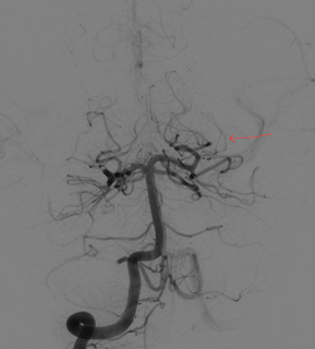
- Squiggly red line : approx start of cavernous sinus/cavernous segment
- Dorsal (or inferior) ICA aneurysm -- not quite SHA, too distal, one would expect a SHA aneurysm to be more proximal. SHA aneurysms point medially and inferiorly
- There are some people who believe that more tortuous carotid siphon = increased likelihood of forming ICA aneurysms
- There is also a pericallosal aneurysm -- kind of weird anatomy. There are 2 pericallosal arteries and they come of ACA really late.


Friday, November 6, 2015

HYPOTHALAMIC LESIONS

All images from this paper: http://www.ncbi.nlm.nih.gov/pubmed/17620469

Embryology: anterior pit from rathke's pouch (from mouth), posterior + infundibulum from neuroectoderm

Lesions:

Craniopharyngioma: 
adamantinomatous - childhood variant, very cystic, very heterogenous 

papillary - adult variant, smaller, more homogenous 

Germinoma

young kids, can have pituitary/pineal synchronous lesions, homogenous, enhancing, non-cystic/heterogenous 

Hamartoma

older kids, tuber cinereum, can be pedunculated or sessile (more likely to be assoc with gelastic seizures & precocious puberty if latter). These do NOT enhance. 

Dermoid Cyst
 well-circumscribed, ectodermal cysts of fat/sebaceous tissue/hair/etc. Typically T1 bright and suppress on STIR, but not as bright and not as suppressy as lipomas. Typically occur in the midline although not commonly at suprasellar. rarely enhance or have calcifications
Vs epidermoid cysts, which have the MRI appearance of dense CSF, more commonly appear off-center - esp parasellar 
Vs arachnoid cysts which look just like CSF because they are 

Rathke's Cleft Cyst
 well circumscribed, midline, between A&P pituitary, typically do not enhance/have calcifications

Hypothalamic/Chiasmatic glioma: 
 very often NF1 associated. larger tumors = more heterogenous, enhancing. 

Ganglioglioma
 very rare, well circumscribed, can be cystic, slow growing. 

Encephalitis 
 usually viral, with clinical evidence of infection 

LCH 
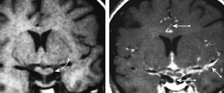
LCH - has unexplained predilection for pituitary stalk and infundibulum. hypothalamic lesions enhance. 

Sarcoid

nodular thickening of chiasm, stalk, infundiblum from granulomatous involvement of dura. predilection for skull base -- neurosarcoid often manifests around hypothal/pit. meningeal enhancement. can invade virchow-robin spaces.