Wednesday, March 11, 2015

Neurofibroma 

General:
- neurofibromas are also a neoplastic proliferation of schwann cells, but the tumors are more heterogenous -- contain fibroblasts, perineural cells, residual axons
- while schwannomas tend to be well circumscribed tumors that push nerves to the side, neurofibromas are non-encapsulated and more invasive
- For more details see blog post from oct 8, 2014
- Associated with NF1 - in these patients, can transform to MPNST

Variants:
- localized soft tissue tumor
- diffuse neurofibroma can grow into large lesion
- plexiform variant - expands nerve fascicles

Path:
(from Dr.Pytel's CNS tumors lecture)
- abundant wavy collagen deposits, "carrot shavings" 

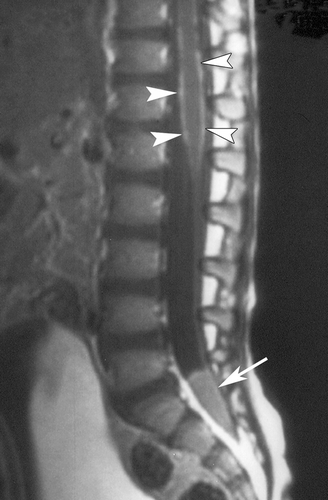
On imaging, can be virtually indistinguishable from schwannoma


MPNST 

General:
- may look like high grade sarcoma
- 50% arise from plexiform neurofibroma in pt with NF1
- 5% of NF patients develop a MPNST

Path:


Many nuclei, cellular spindle cell tumor, lots of mitotic activity (contrast with path image above)
source: Dr.Pytel's lecture

Tuesday, March 10, 2015

Schwannoma 

General
- Peripheral nerve sheath tumors
- 8th nerve typically vestibular branch (rarely other CN), nerve roots, peripheral nerves
- Other nerve sheath tumors: neurofibromas, MPNST
- Bilateral = NF2
- Rarely undergo malignant transformation

Path
- Gross: well-circumscribed, extra-axial
- Source: Dr.Pytel's CNS tumors lecture


Antoni A and antoni B


Verucay bodies - cells without nuclei


Imaging:

acoustic


source: radiopaedia

source: headneckbrainspine

Monday, March 9, 2015

Medulloblastoma 

General
- from cells resembling immature neurons of cerebellum
- Most common malignant/high grade tumor of children - also most common posterior fossa tumor of children (30-40%)
- Mean age of appearance - 9 years. When they occur in adults (rare) typically 3rd-4th decade, better prognosis
- May recur locally or spread in CSF - drop mets in spinal cord, or explants in hemispheres.

Prognosis
- Heterogenous group of tumors - prognosis varies widely depending on molecular signaling.
- Tx: surgical resection, chemo, whole brain and spine RT to manage potential mets/prevent spread.
- 5-year survival 50-70%, but with high morbidity 2/2 RT - i.e. short stature due to hypothalamic and spinal bony RT, secondary malignancy.

Path
- small blue cell tumor - sheets of undifferentiated cells. Sometimes rosettes.

- "rosettes" 

- sheets of blue cells

Neuronal lineage - stain + with neuronal markers like synaptophysin and NeuN (nuclear marker of neurons)

Imaging:
(source: radiopaedia, this paper)
- CT bright - may have significant cystic/calcific components
- T1 dark
- 90% enhance, usually heterogenously

- medullo with leptomeningeal spread along anterior pons 

- classic medullo - emerging from cb vermis 

- leptomeningeal mets. 

Sunday, March 8, 2015

CNS Lymphoma

General
- Primary Sporadic extranodal lymphoma (older patients) - usually EBV-neg - no extra-CNS disease
- Primary CNS lymphoma of immunocompromised patients - can occur at any age - usually EBV-associated
- Secondary CNS lymphoma = mets from primary lymphoma elsewhere, most common - 2/3 leptomeningeal/subependymal disease
- Single/multiple lesions, or diffuse infiltrate
- Mostly large B-cell lymphomas
- Tx with intrathecal chemo and RT
- Poor prognosis


Path:
(source: Dr.Pytel's CNS tumors lecture)
- no processes emanating from cells


Imaging: 
- Data and images sourced from this AJNR paper
- Immunocompetent: usually homogenously enhancing (90%), ring-enhancing (0-13%)
- AIDS patients - heterogenously enhancing 75% of the time
- homogenously enhancing, can be dural-based 

- AIDS related disease - can be heterogenous, ring-enhancing 


Friday, March 6, 2015

Oligodendroglioma 

General:
- Adult tumor
- poorly circumscribed, parenchymal, infiltrating growth pattern
- WHO grade II (standard)
- WHO grade III (anaplastic) - more mitotically active
- If there is necrosis, it is considered a GBM
- Better prognosis than infiltrating astrocytomas - may be because they respond better to therapy
- Can occur anywhere in the brain - including intraventricularly (may resemble central neurocytoma) 

Molecular biology: 
- 1p and 19q codeletion - marker of response to chemotherapy and radiation and thus better prognosis. More characteristic of oligodendroglioma (vs astrocytoma) 
- Intact 1p/19q: worse response to chemo/RT


Path 
- fried egg appearance - round nuclei with halos (retraction artifact?)
- in background of normal neurons - infiltrating cells
- can occur with fine/small branching capillaries giving it a "chicken wire" appearance

Imaging: 
- Usually calcified (70-90% calcified)
- Sometimes focally hemorrhagic. Sometimes (20%) cystic.
- CT bright: calcifications or hemorrhage, may erode overlying skull 
- T1 dark, T2 bright 
- Minimal peritumoral edema
- May enhance, enhancement usually heterogenous 
- These tumors typically go all the way to the outer cortex of the brain 

Thursday, March 5, 2015

GBM

Occurs in adults 
- Mean survival ~1 year (vs 3 years for AA, 6-8 years for grade II astrocytoma) 
- Can occur spontaneously as a primary tumor (EGFR, PTEN, p16) 
- Can evolve from grade II/III tumors (p53, IDH-1/IDH-2) IDH changes methylation of genes, changes expression 

Subtypes: 
- may arise from specific subtypes of cells? 


Path: 
- palisading cells around central areas of necrosis 

- endovascular cell proliferation - plump endovascular cells proliferating out of proportion - almost appear like glomeruli 

- giant cell variant 

Imaging: 
- heterogenous appearing 
- usually contrast-enhancing, sometimes ring-enhancing 
- can be bilateral or multifocal 


- butterfly GBM

- multifocal GBM 

Wednesday, March 4, 2015

Low grade astrocytoma: 

Occurs in adults
- Mean survival 6-8 years
- Differentiate from Anaplastic (who III), which has more anaplasia and more mitoses, and GBM which has tumor necrosis and endothelial proliferation
- Can transform into higher grade astrocytomas over time.

Gross path:
- No discrete circumscribed lesion
- Diffuse expansion of the brain parenchyma.

Path:
- Increased cellularity of brain 
- Ovoid to elongated nuclei, hyperchromasia 

- gemistocytes - pink inclusions around nuclei, indicate reactive astrocyte
- must have > 20% gemistocytes to be considered a "gemistocytic" tumor (oligos and others can have occasional gemistocytes)
- worse prognosis than WHO-grade matched controls

T1 dark, T2 bright, non-enhancing
source: headneckbrainspine.com

Tuesday, March 3, 2015

Pilocytic astrocytoma: 

- well circumscribed, often cyst with mural nodule 
- Usually peds 
- Cerebellum, hypothalamus, optic nerve (in NF-1) 
- WHO grade I 
- Prognosis varies greatly depending on resectability, which depends on location
- Biologically distinct from the diffuse/infiltrating astrocytomas (grade II, III, IV)
- 17q loss, BRAF mutations

- Path 

- variation between loose pale areas, dense pink areas. 

- bipolar spindled cells with hair-like processes. fibrillary background 
- Rosenthal fibers - glassy pink deposits of GFAP, a protein produced by astrocytes 

Imaging: 

hypothalamus

optic nerve 


classic cerebellar 

Monday, March 2, 2015

Meningioma

Arise from arachnoidal cells
Well circumscribed, vascular tumors
Dural based 

Path: (all images from Dr.Pytel's CNS tumors lecture)
lobulated, "islands of cells" separated

whorls, relatively a-cellular/not dense in cells, with normal appearing cells that all look the same.

Pseudoinclusions, monomorphic ovoid cells. 

Subtypes of meningoma: 

Signs of poor prognosis/higher grade: 
- Tumor invading brain tissue 
- Proliferative activity (MIB-1) staining
- Lots of mitotic activity 

Prognosis: 

WHO grade I: recurrence 7-20%
WHO grade II (atypical): recurrence 29-40%
WHO grade III (anaplastic): recurrence 50-78%

Female predominance, esp in spine meningomas - 10:1 ratio